Please use Microsoft Edge, Google Chrome or [Firefox](https://getfirefox. com/).

CENTRO OCULISTICO BESSO Sanchez Lasa Enrique (Lugano)
Do you own or work for CENTRO OCULISTICO BESSO Sanchez Lasa Enrique?
About Us
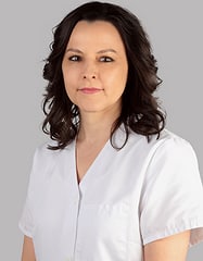
ENRIQUE SANCHEZ LASA
Med. Spec. FMH Ophthalmology and Ophthalmic Surgery
INTRODUCTION:
Modern ophthalmology is one of the most high-tech specialties, requiring constant updates and investment on the part of the physician.
Regular checkups by his ophthalmologist allow early diagnosis of diseases, diseases that if not caught in time can sometimes lead to blindness.
The Besso eye care center in Lugano as well as the medical office in Biasca offer a broad spectrum of diagnostics and treatments across the board.
Our technical services are as follows:
1. Autorefractometry: a method with infrared technology, harmless and accurate for assessing visual defects (hyperopia - myopia - astigmatism).
It helps to determine the dioptric power of glasses or contact lenses, which will be prescribed by the eye doctor.
After instillation of drops to temporarily block accommodation (cyclopegic examination), even more accurate measurements are obtained in children.
2. The portable Spot-Vision instrument (Watch Allyn): allows the same measurements at a distance of about 1 meter. It is very useful particularly in young children (under 4 years of age), where classic examination with the autorefractometer may prove difficult to achieve. It allows early diagnosis of vision defects and a detailed written report for the pediatrician.
3. Frontifocometer or device for measuring the dioptic power of eyeglasses (or contact lenses).
4. Phoropter: allows a computerized examination of vision, giving the correction for prescribing glasses or contact lenses
5. Tonometry or measurement of intraocular pressure:
1. Non-contact tonometry:
(a) Air-blow tonometer: allows for quick screening and can be performed by the medical office assistant.
2. Contact tonometry or corneal applanation, after instillation of anesthetic eye drops:
(a) Examination performed at the fissure lamp, after instillation of anesthetic eye drops..:
1. Goldmann tonometry
2. resonance sensor tonometry: Bioresonator®
b). Tonometry with a handheld instrument, the Tonopen®: for children, for bedridden patients or in home visits
6. Slit lamp examination (LAF): slit lamp examination (LAF) or biomicroscopy by means of a slit of light, allows different ocular structures to be seen:
(a) anterior segment: eyelids, conjunctiva, tear film, cornea, anterior chamber, iris-pupil and crystalline lens.
(b) Posterior segment: vitreous, retina with its vessels and optic nerve
7. Corneal topography: noninvasive qualitative and quantitative examination of the cornea using a videokeratograph or a 360° Scheimpflug camera. It allows measurements of the overall dioptric power of the cornea as well as the study of the anterior and posterior curvatures of the cornea. A corneal thickness map or topography (pachymetry) is also obtained.
Corneal topography allows early diagnosis of diseases of the corneal surface or stroma, such as keratoconus.
It is performed as a preoperative examination of suitability for excimer laser vision defect correction as a follow up after excimer laser treatments (PRK = photorefractive keratectomy or LASIK = laser in situ keratomileusis).
The topograph Sirius® designs 22 rings and analyzes more than 100,000 points in a corneal diameter up to more than 12 mm in diameter. It also allows tear film analysis (bucket eye, contact lenses) and with the Scheimpflug chamber allows visualization of the cornea, irido-corneal angle and lens.
8. ** Biometry: is the biometric study of the eye with measurements of ocular structures.
The classical contact ultrasound probe method after instillation of anesthetic eye drops is painless and harmless (ultrasound).
A more modern or non-contact method, is automatic with infrared technology and optical coherence inteferometry is optical biometry (devices such as Aladdin®, Lensar®, IOLmaster®).
The new technology allows for quick, simple, and touch-free measurements that can be delegated to a nonphysician office staff member. In certain cases, particularly when the cataract is very dense one must still resort to the use of the classical ultrasound method.
The Aladdin® device: allows measurements of the axial lengths of the eye and corneal curvature values (keratometry), measurements that are indispensable for calculating the intraocular lens (IOL), which will be implanted during cataract surgery. The device simultaneously performs a study of the ocular surface (topography), an analysis of the optical aberration of the eye (aberrometry), and the study of the pupil in both bright and dark-adapted conditions, thus allowing the best choice of the type of IOL to be inserted in cataract surgery.
9. Corneal specular microscopy (Topcon SP-1p): this SP-1p instrument from the Topcon company analyzes central corneal thickness or pachymetry, visualizes endothelial cells with cell count and study of cell morphology. It allows diagnosis of problems of the cornea, particularly the endothelium. The examination is important both before intraocular surgery as well as in postoperative follow-up, particularly useful for cataract surgery or in corneal surgery (transplants).
10. GDX-NFA: confocal laser scanning technology that allows indirect measurement of the optic nerve and its nerve fibers.
Being a noninvasive examination, it is easy to perform and allows along with the realization of visual fields (computerized campimetry), the tracking of clinical evolution in cases glaucoma.
It can be observed in the evolution chart of the different examinations, visualizing over time any worsening that motivates a change in therapeutic attitude (eye drops, laser or surgery)
11. OCT (Optical Coherence Tomography): is a fast and harmless examination that allows virtual cuts of the desired area of the eye.
The device records 70'000 images per second allowing fast and accurate acquisition of the analyzed structures with excellent resolution (2.5ų).
OCT is useful in diagnosing the various forms of macular degeneration, dry form and wet form with exudation of intra- or sub-retinal fluid. It has enabled better understanding of the disease, earlier diagnosis and effective treatment and follow-up of maculopathy.
Using a special module, for the cornea, OCT examination allows measurement of corneal thickness (pachymetry), differentiating the epithelial layer from the deeper stromal layer. In this sense it is useful in the diagnosis of corneal pathologies and in the follow-up of the patient after laser refractive surgery or treatment of keratoconus with Cross-linking.
Finally, it allows the study of the entire anterior part of the eye, with measurements of the corneal irido angle, position of the natural or artificial lens (IOL).
OCT also reveals a detailed analysis of optic nerve anatomy and nerve fiber study. It is part of the patient's routine examination for both diagnostic and follow-up glaucoma.
AngioOCT is a breakthrough in the field of ophthalmology by facilitating diagnostics in diseases of the macula and retina: for example:
- macular edema
- diabetic retinopathy
- age-related macular degeneration
- central serous coiriopathy
- presence of sub- or intra-retinal neovascularization with quantification of fluid presence in age-related macular degeneration or even maculopathy in severe myopia.
- Status after vascular venous thrombosis.
Allows evaluation of the outcome after intravitreal injection treatments with drugs such as Anti-VEGF (Lucentis, Eylea, Avastin,) or intravitreal injections of corticoids (Ozurdex) or other drugs, (e.g., Jetra to make a vitreo-retinal traction dissolve).
The advantage of AngioOCT over conventional angiography using contrast agents (such as; Fluorescein, Indocyanine green) is its safety without side effects related to the use of dye products injected into the patient.
It can be performed in general without dilatation, it is a fast examination (about 5 minutes), in total safety it is feasible in all types of patients especially in pregnant woman, patients with renal problems or major cardio-respiratory risk without forgetting patients who may be allergic to the contrast medium.
12. Fundus photography and angiography (Fluorescein): the digital retinal camera allows:
- color images without dilation (45°) or after dilation retinal image to (50°),
with visualization of the retina, retinal circulation and optic nerve
The use of a special filter also enables particularly effective evaluation of nerve fibers (glaucoma); another filter enables auto-fluorescence imaging of the macula, thus without the use of contrast medium, which is interesting in macular diseases such as macular dystrophies or age-related macular degeneration.
Sometimes a dye or contrast agent (Fluorescein) injected diluted into the vein is used to allow better visibility of retinal pathology; this examination requires patient consent and is performed under the supervision of a physician-anesthesiologist.
13. CAMPIMETRY
A) Kinetic fieldimetry: allows assessment of the visual field by a light spot that varies in intensity and size and moves from the periphery toward the center. A simple to perform and quick examination, particularly useful for neuro-of-tamological disorders.
B) Computerized campimetry: the patient fixes a central point and light stimuli are emitted, of varying luminous intensity so as to determine the discrimination threshold of the light point, in this way a representation of the visual field is obtained for each eye.
Octopus 900: allows a 90° study for each eye, has a specific program for driving ability and another program to show the field of view defect particularly related to eyelid pathologies (budget before upper blepharoplasty surgery).
Octopus 600: allows a central 60° study for each eye and presents as a special feature a "Pulsar "perimetry program that by simultaneously evaluating contrast sensitivity and brightness of stimuli, allows earlier diagnostics of glaucoma.
14. Othoptic examination with Hess-weiss table: allows evaluation of oculo-motor disorders to the projection of a light on a Hess-weiss board. Useful for determining oculo-motor paralysis, congenital strabismus acquired for example after trauma (fracture of the orbit), vascular accident, diabetes or inflammation of the central nervous system
15. Color test:
- (24 tables):
- Farnsworth test (15 or 100 round color tablets).
Allows the diagnostic of congenital abnormalities of color perception such as color blindness (green-red axis or blue-yellow axis) or acquired abnormalities such as macular or optic nerve pathologies, (often happens after inflammation or taking medications).
16. Stereoscopic vision examination: tests our ability to perceive visually in three dimensions, or lack thereof, by a simple test (Lang's test or with polarized filters).
This test is important for driving ability.
17. Laser treatments: in the modern therapeutic arsenal available to the ophthalmologist, different types of lasers are an important part.
They allow treatment of corneal curvature abnormalities or corneal scarring by Excimer laser. Posterior capsulotomy or cleaning of the la capsule (secondary cataract), again allows the patient to see well after this treatment performed by Yag laser.
In narrow-angle glaucoma, peripheral laser iridotomy allows bypass and improves fluid circulation between the anterior and posterior chambers, lowering intraocular pressure.
The laser is also very useful in performing a trabuloplasty or cleaning of the trabeculum, which is the natural filter and greatest point of resistance to fluid leaving the eye. In the case of glaucoma, the trabeculum in general is pathological, "dirty," with dead, inflammatory cells or pigment, which go to close the trabeculum. Particularly useful today is selective trabeculoplasty with laser-YAG (SLT), which does not damage the eye and can be repeated at will.
In the glaucoma setting, a probe with external application to the limbus is also used to selectively target the aqueous production site, called the ciliary body. Cyclodestruction with an 810 nm diode laser is then performed.
Another application of the laser is the realization of a barrier with Argon laser, which allows treating a retinal hole and generally preventing retinal detachment.
The different platforms available from us are as follows:
1. Lumenis Novus Spectra (Multiple platform with 3 lasers): Argon laser, YAG laser, and SLT laser.
2. Quante Medical Argon 532 nanometer laser: useful for retinal photocoagulation treatments.
3. Quante Medical Argon 1064 nanometer laser: useful for peripheral iridotomy and capsulotomy (secondary cataracts)
4. Iridex Diode 810 nanometer laser: of retinal photocoagulation and tumor treatment with TTT (trans pupillar Thermo-Therapy), with external probe for cyclodestruction in case of neovascular glaucoma or resistant to all other therapies
5. Laser Excimer Amaris 500: laser working very fast at 500 Herz, allows correction of all types of vision defects (hypermetropia-astigmatism-myopia), combined treatments and as well as treatment of presbyopia thanks to a special module the Presby-Lasik.
18. Module of eye surgeries:
1. Extraocular surgery with glasses or telelopes:
- eyelid surgery to improve the functional or cosmetic appearance of the eyelids in
conditions such as chalazio, ectropion, ptosis, blepharocalasia, etc.
2. General ocular surface surgery with an operating microscope: in cases of
pinguecula, pterygium, corneal deposits (such as keratopathy, etc...)
3. Intraocular surgery:
(a) cataract: replacement of an opaque lens with an intraocular lens.
(b) glaucoma: nonperforating filtering surgery such as deep sclerotomy or
viscocanulostomy,
glaucoma: use of valves or drainage tubes (e.g. Baerveldt tube or tube
Ex-Press)
Minimally invasive surgery on the trabeculate: ei-stent or direct subconjunctival bypass (Xen implant)
(c) Intravitreal injection: for treatment of macular degeneration, diabetic macular edema, after venous thrombosis or macular vitreous traction (Avastin, Eylea, Lucentis, Jetrea, Ozurdex...
-
Excimer lasers: for correction of vision defects
-
Transepithelial cross-linking: to stabilize the cornea (keratoconus)
19. BeatyCell aesthetic treatments
1. Botulinum Toxin A (Azzalure®, Vistabel®): botulinum toxin is used to treat wrinkles due to facial expression or facial mimicry wrinkles particularly present in the upper half of the face.
Effectively, the toxin prevents muscle contraction by inhibition of nerve transmission at the level of the neuronal plate. The effect begins to be seen after 4-5 days and lasts about 4 months. Used as early as the 1970s to treat blepharospasm and strabismus in the field of ophthalmology, botulinum toxin was later used in the neurological field to treat complex facial spasms. For about 20 years it has been used in the field of aesthetic medicine; botulinum toxin is a poison and if used appropriately can be found to be dangerous and lead the patient to death.
It is very important to go to an experienced medical specialist and if possible registered in national or international societies of aesthetic medicine.
The cost of the treatment ranges from CHF 150 to CHF 600 per session depending on the area and the amount of product used
2. Filler or skin filling: with a viscoelastic material, Ac. Hyaluronic (Surgidem®, Juvederm®), which is used with different concentrations and molecular weight depending on the areas and wrinkles to be treated, generally lower half of the face. Hyaluronic acid has three properties: 1. allows a filling by giving volume and thus decreasing wrinkles. 2. it moisturizes up to 1000 times its molecular weight, thus giving intense hydration to the skin with consecutive cunateous smoothing. 3. helps one's skin regenerate with stimulation of fibroblasts, cells that produce collagen and elasticity, thus improving visual appearance and skin elasticity.
The treatment costs CHF 300 to CHF 400 per vial depending on the product and amount used.
3. Superficial or medium-depth peeling: depending on skin condition. Allows superficial wrinkles to be corrected, spots or pigmentation irregularities to be diminished, and finally allows better regulation of skin seborrheic secretion. It acts as an exfoliant of the old superficial state and acts deeper to stimulate the skin (neocollagen) and decrease skin pigmentation with action on snow cells. It is a painless treatment and lasts about 10 minutes and can be repeated as needed.
Contraindications: Herpes, collagen diseases and pregnancy.
The cost of peeling ranges from CHF 50 to CHF 150 per session.
4. Other cosmetic treatments can be performed in our beauty center Beauty cell
This text has been machine translated.
Location and contact
CENTRO OCULISTICO BESSO Sanchez Lasa Enrique
-
office address
Via Besso 31 6900 Lugano
-
Phone
0919... Show number 091 950 93 55
-
Write an e-mail
- Visit site Visit site
reviews
Do you wish to rate "CENTRO OCULISTICO BESSO Sanchez Lasa Enrique"?

There are no reviews for this company yet.
Have you any experience of this company?

* These texts have been automatically translated.
Other listers

CENTRO OCULISTICO BESSO Sanchez Lasa Enrique


